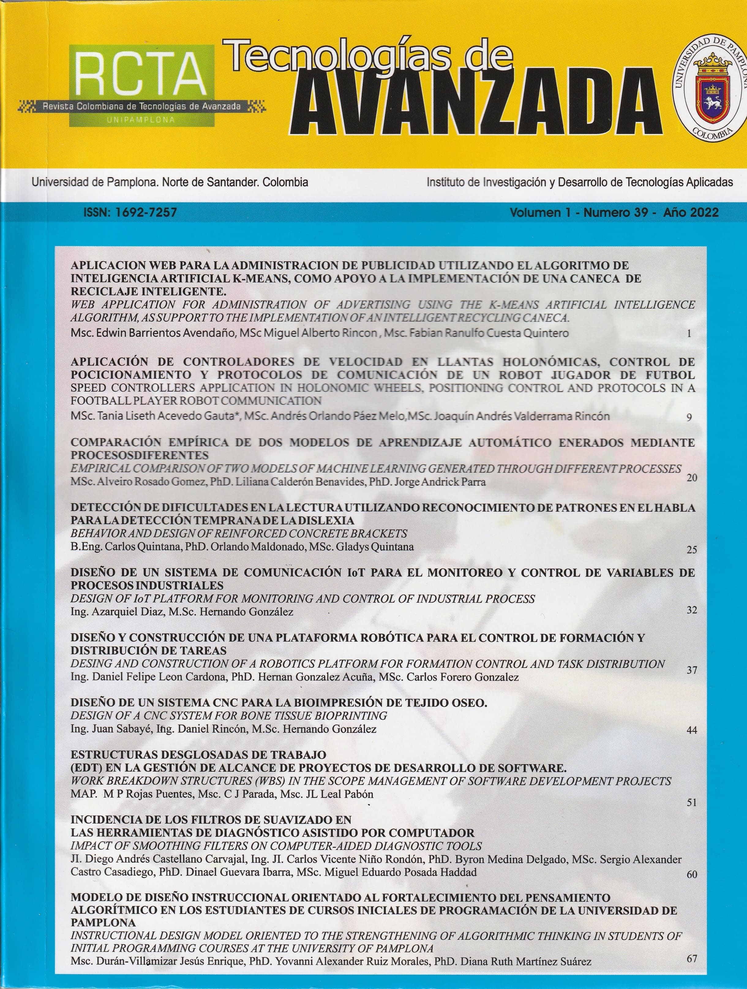
Incidencia de los filtros de suavizado en las herramientas de diagnóstico asistido por computador
DOI:
https://doi.org/10.24054/rcta.v1i39.1376Palabras clave:
Procesamiento de imágenes médicas, filtros de suavizado, lesiones cutáneas, correlaciónResumen
El procesamiento de la imagen en el campo biomédico hace referencia a la etapa inicial orientada en mejorar la calidad de las imágenes, eliminando los ruidos irrelevantes y secciones no deseadas en el fondo de las imágenes. En este documento se evalúa la incidencia de los filtros de suavizado (filtro bilateral para conservación de bordes, filtro de mediana y el filtro gaussiano) en el procesamiento digital de imágenes médicas dermoscópicas como apoyo a las herramientas de diagnóstico asistido por computador. El método fue probado con imágenes del Dataset HAM10000, compuesto por imágenes de lesiones cutáneas pigmentadas. Para el procesamiento digital se utilizaron herramientas basadas en código abierto como lenguaje Python y la librería especializada en visión por computador OpenCV. La validación se realizó por el método de correlación entre la imagen original en escala de grises y la imagen filtrada por cada uno de los filtros de suavizado, obteniendo un porcentaje en relación de cambio medio de 99.460 % para el filtro bilateral,
99.396 % para el filtro gaussiano, y 99.335 % para el filtro de mediana.
Descargas
Referencias
Al-abayechi, A. A. A., & Abu-Almash, F. S. (2020). Skin Lesion Border Detection Based on Best Statistical Model Using Optimal Colour Channel. Journal of Autonomous Intelligence, 3(1), 26. https://doi.org/10.32629/JAI.V3I1.131
Asokan, A., & Anitha, J. (2020). Adaptive Cuckoo Search based optimal bilateral filtering for denoising of satellite images. ISA Transactions, 100, 308–321. https://doi.org/10.1016/J.ISATRA.2019.11. 008
Castellanos, W. A., Suarez, O. J., & Garcia, A. P. (2018). Usability in virtual learning environments, an approach to the integrated grid (IG) application. Paper presented at the Proceedings of the LACCEI International Multi-Conference for Engineering, Education and Technology, , 2018-July doi:10.18687/LACCEI2018.1.1.497
Garg, B., & Sharma, G. K. (2016). A quality- aware Energy-scalable Gaussian Smoothing Filter for image processing applications. Microprocessors and Microsystems, 45, 1–9. https://doi.org/10.1016/J.MICPRO.2016.02.012
Garcia, A. P., Suarez, O., & Castellanos, W. (2016). ERAAE virtual library. CHILECON 2015 - 2015 IEEE Chilean Conference on Electrical, Electronics Engineering, Information and Communication Technologies, Proceedings of IEEE Chilecon 2015, 911-916. doi:10.1109/Chilecon.2015.7404681
Huang, H.-W., Hsu, B. W.-Y., Lee, C.-H., & Tseng, V. S. (2021). Development of a light-weight deep learning model for cloud applications and remote diagnosis of skin cancers. The Journal of Dermatology, 48(3), 310–316. https://doi.org/10.1111/1346-8138.15683 Ibrahim, E., Ewees, A. A., & Eisa, M. (2020).
Proposed Method for Segmenting Skin Lesions Images. Lecture Notes in Electrical Engineering, 569, 13–23. https://doi.org/10.1007/978-981-13-8942-9_2
Kang, X., Zhang, X., Li, S., Li, K., Li, J., & Benediktsson, J. A. (2017). Hyperspectral Anomaly Detection with Attribute and Edge-Preserving Filters. IEEE Transactions on Geoscience and Remote Sensing, 55(10), 5600–5611. https://doi.org/10.1109/TGRS.2017.27101 45
Lynn, N. C., & Kyu, Z. M. (2018). Segmentation and classification of skin cancer Melanoma from skin lesion images. Parallel and Distributed Computing, Applications and Technologies, PDCAT Proceedings, 117– 122. https://doi.org/10.1109/PDCAT.2017.0002 8
Mane, S., & Shinde, S. (2018). A Method for Melanoma Skin Cancer Detection Using Dermoscopy Images. 4th International Conference on Computing, Communication Control and Automation, ICCUBEA 2018. https://doi.org/10.1109/ICCUBEA.2018.86 97804
Márquez Díaz, J. E. (2020). Deep Artificial Vision Applied to the Early Identification of Non-Melanoma Cancer and Actinic Keratosis. Computación y Sistemas, 24(2), 751–766. https://doi.org/10.13053/cys-24-2-2901
Neshatpour, K., Koohi, A., Farahmand, F., Joshi, R., Rafatirad, S., Sasan, A., & Homayoun, H. (2016). Big biomedical image processing hardware acceleration: A case study for K- means and image filtering. Proceedings - IEEE International Symposium on Circuits and Systems, 1134–1137. https://doi.org/10.1109/ISCAS.2016.75274 45
Ottom, M. A. (2019). Convolutional neural network for diagnosing skin cancer. International Journal of Advanced Computer Science and Applications, 10(7), 333–338. https://doi.org/10.14569/IJACSA.2019.010 0746
Padmavathi, K., & Thangadurai, K. (2016). Implementation of RGB and Grayscale Images in Plant Leaves Disease Detection – Comparative Study. Indian Journal of Science and Technology, 9(6), 1–6. https://doi.org/10.17485/IJST/2016/V9I6/7 7739
Singhal, P., Verma, A., & Garg, A. (2017). A study in finding effectiveness of Gaussian blur filter over bilateral filter in natural scenes for graph based image segmentation. 4th International Conference on Advanced Computing and Communication Systems, ICACCS 2017,4–9. https://doi.org/10.1109/ICACCS.2017.8014612
Xu, Z., Sheykhahmad, F. R., Ghadimi, N., & Razmjooy, N. (2020). Computer-aided diagnosis of skin cancer based on soft computing techniques. Open Medicine (Poland), 15(1), 860–871. https://doi.org/10.1515/MED-2020- 0131/MACHINEREADABLECITATION/ RIS
Zhu, F., Liang, Z., Jia, X., Zhang, L., & Yu, Y. (2019). A Benchmark for Edge-Preserving Image Smoothing. IEEE Transactions on Image Processing, 28(7), 3556–3570. https://doi.org/10.1109/TIP.2019.290877
Descargas
Publicado
Número
Sección
Licencia
Derechos de autor 2022 Sergio Alexander Castro Casadiego, Diego Andrés Castellano Carvajal, Carlos Vicente Niño Rondón, Byron Medina Delgado, Dinael Guevara Ibarra, Miguel Eduardo Posada Haddad

Esta obra está bajo una licencia internacional Creative Commons Atribución-NoComercial 4.0.


